Unused files
From Slicer Wiki
The following files exist but are not embedded in any page. Please note that other web sites may link to a file with a direct URL, and so may still be listed here despite being in active use.
Showing below up to 500 results in range #1 to #500.
View (previous 500 | next 500) (20 | 50 | 100 | 250 | 500)
 3DSlicerLogo.png 424 × 236; 35 KB
 3DSlicer2.jpg 175 × 175; 8 KB
 LogoNCIA.jpg 500 × 53; 11 KB
 SlicerTraining5 fMRI.pdf 1,650 × 1,275, 109 pages; 4.67 MB
 Slicer 2.6 page EMAtlasBrainClassifier.pdf 1,650 × 1,275, 62 pages; 1.89 MB
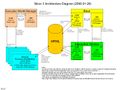
 2007 Slicer Architecture slides.ppt ; 321 KB
 MRML Overview for Slicer.ppt 1,600 × 1,200; 261 KB
 2007 EMSegmenterTutorial.pdf 1,275 × 1,650, 23 pages; 3.33 MB
 SavingSlicerData.pdf 1,650 × 1,275, 43 pages; 1.19 MB
 Logo-x.jpg 54 × 61; 1 KB
 Logo-ncrr.jpg 232 × 50; 4 KB
 Logo-nibib.gif 170 × 130; 8 KB
 Logo-dod.jpg 622 × 62; 18 KB
 Logo-newdod.jpg 280 × 62; 8 KB
 Logo-birn.jpg 770 × 50; 73 KB
 Logo-cimit.jpg 188 × 100; 7 KB
 Logo-newbirn.jpg 207 × 50; 7 KB
 Logo-spl.jpg 150 × 120; 4 KB
 Logo-csail.gif 150 × 115; 5 KB
 Logo-isomics.gif 252 × 194; 10 KB
 Logo-kitware.gif 300 × 49; 6 KB
 Logo-nmr.gif 319 × 74; 5 KB
 Logo-newnih.jpg 165 × 118; 4 KB
 Logo-isomics2.gif 189 × 117; 7 KB
 Logo-newmartinos.gif 240 × 67; 3 KB
 Logo-nih2.jpg 84 × 94; 2 KB
 Logo-nacnew.jpg 88 × 156; 2 KB
 Logo-nmr2.gif 62 × 66; 1 KB
 Logo-nmr2 2.gif 62 × 58; 2 KB
 Logo-Martinos2.gif 240 × 67; 3 KB
 Logo-nihlogo.jpg 54 × 61; 1 KB
 Logo-hcnr.gif 312 × 87; 25 KB
 Logo-hcnr-mj.gif 312 × 87; 25 KB
 Logo-dodnew.gif 189 × 62; 6 KB
 Logo-nibibnew.gif 128 × 98; 4 KB
 Logo-namicnew.jpg 126 × 143; 9 KB
 Logo-nacnew2.jpg 88 × 156; 5 KB
 Logo-hcnrnew.gif 130 × 77; 9 KB
 Logo-isomicsnew.gif 142 × 88; 5 KB
 Logo-csailnew.gif 113 × 87; 3 KB
 Logo-namicNEW.jpg 126 × 143; 9 KB
 Tatrc.gif 120 × 73; 3 KB
 Nih-logo.gif 109 × 100; 2 KB
 Logo-csail.jpg 113 × 87; 3 KB
 Logo-csailnew.jpg 150 × 115; 5 KB
 Logo-csailNEW.jpg 150 × 115; 5 KB
 Logo-csailv2.jpg 113 × 87; 3 KB
 Logo-NIBIB.gif 54 × 66; 2 KB
 Logo-HCNR.gif 65 × 87; 5 KB
 Logo-ncrrblack.jpg 2,584 × 638; 171 KB
 Logo-NMRMGH.gif 624 × 668; 21 KB
 Logo-NMR.gif 624 × 668; 21 KB
 Logo-NMR2.gif 624 × 668; 21 KB
 TATRC.gif 166 × 99; 4 KB
 Logo-CSAIL.jpg 106 × 82; 2 KB
 Logo-CSAIL2.jpg 96 × 83; 2 KB
 Logo-ISOMICS.gif 140 × 39; 2 KB
 Logo-SURGPL.jpg 150 × 120; 4 KB
 Archip 2007 Neuroimage.gif 497 × 447; 290 KB
 Logo-NIBIB.jpg 553 × 682; 62 KB
 Logo-NIBIB2.jpg 553 × 682; 62 KB
 Logo-Nibib.jpg 553 × 682; 62 KB
 Nibib.jpg 553 × 682; 62 KB
 Fedorov-Supercomputing2006-fig3.png 1,534 × 537; 1.05 MB
 3DSlicerIcon32x32.png 32 × 32; 2 KB
 3DSlicerLogoIcon32x32x256.gif 32 × 32; 2 KB
 Li-ClinicalAnatomy2006-2-fig-mj.png 1,533 × 498; 655 KB
 Margulies-AJOG2007.pdf 1,200 × 1,612, 5 pages; 475 KB
 Logo-pnl.jpg 191 × 106; 11 KB
 Logo pnl2.jpg 168 × 182; 55 KB
 Logo pnl.png 168 × 182; 55 KB
 Logo pnl.jpg 173 × 190; 41 KB
 SanJose-CAS2007.pdf 1,275 × 1,650, 14 pages; 864 KB
 Snowflakes.gif 198 × 203; 11 KB
 Dlpfc outlined for jim.png 717 × 635; 338 KB
 WM color.png 1,362 × 1,126; 554 KB
- Example.swf ; 26 KB
 SmallEmbodiment.png 600 × 464; 83 KB
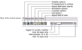
 SliceGUI.png 1,032 × 419; 150 KB
 SliceControllerGUI.png 589 × 360; 20 KB
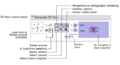 ViewControllerGUI.png 592 × 319; 25 KB
 VolumesModule.png 403 × 1,057; 20 KB
- VR Load Basics.swf ; 5.63 MB
- VR Threshold.swf ; 8.54 MB
- VR Performance.swf ; 5.65 MB
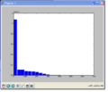 Pythonhist.png 681 × 583; 29 KB
 Tracts.jpg 1,000 × 750; 394 KB
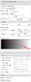
 Diffusion Editor v1.jpg 389 × 582; 196 KB
 DiffusionEditor v7.jpg 390 × 560; 192 KB
 Slicer3LogoHorizontalBeta.png 135 × 70; 11 KB
 Slicer3-Architecture-Layers.jpg 1,600 × 1,200; 218 KB
- SlicerHistoricalTimeline.ppt ; 5.13 MB
- Slicer3-ExecutionModelJune2006.ppt ; 292 KB
- Dti glyphs.swf ; 21.72 MB
- EMSegmentTutorial.tgz ; 41.07 MB
- EMSegmentTutorial Slicer3.zip ; 41.23 MB
 EMSegmentSlicer3 Segmentation.png 1,026 × 821; 77 KB
- PythonModules.zip ; 13 KB
- Logo-SCI.tif 754 × 331; 48 KB
 MouseModeIcons.png 236 × 58; 6 KB
 MouseModeMockup.png 720 × 936; 77 KB
 SB1.png 506 × 542; 123 KB
 SB2.png 506 × 542; 159 KB
 SB3.png 505 × 529; 148 KB
 SB4.png 506 × 543; 188 KB
 SB5.png 505 × 543; 189 KB
 SB6.png 506 × 542; 190 KB
 SB8.png 506 × 543; 191 KB
 SB9.png 507 × 556; 191 KB
 SB10.png 506 × 556; 191 KB
 ToolbarMouseManipulate.png 21 × 21; 469 bytes
 ToolbarMouseSelectRegion.png 21 × 21; 304 bytes
 ToolbarMouseLasso.png 21 × 21; 487 bytes
 ToolbarMouseDeselectAll.png 21 × 21; 343 bytes
 ToolbarMousePlace.png 21 × 21; 400 bytes
 ToolbarMouseRotate.png 21 × 21; 604 bytes
 ToolbarMousePan.png 21 × 21; 363 bytes
 ToolbarMouseZoom.png 21 × 21; 402 bytes
 SlicesFitToWindow.png 21 × 21; 3 KB
 SB7.png 506 × 543; 188 KB
 MRI Robot System Diagram2.png 788 × 580; 25 KB
 Larsenetal2007.png 716 × 558; 462 KB
 Chick.png 178 × 233; 31 KB
- DiffEd Tut Load.swf ; 3.69 MB
- DiffEd Tut Gradients.swf ; 6.27 MB
- DiffEd Tut MeasurementFrame.swf ; 3.1 MB
- DiffEd Tut Testing.swf ; 38.83 MB
- SlicerWithCUDAUS New.zip ; 116.46 MB
 Caption1.PNG 852 × 650; 367 KB
 Caption2.PNG 379 × 360; 13 KB
 Caption3.PNG 855 × 294; 22 KB
 Caption1.png 852 × 650; 367 KB
 Caption2.png 379 × 371; 33 KB
 Caption2.jpg 379 × 371; 33 KB
 XNDchoosetags.png 504 × 504; 20 KB
 XNDmanage.png 504 × 504; 20 KB
 XNDresult1.png 826 × 374; 23 KB
 XNDresult2.png 258 × 416; 11 KB
 XNDresult3.png 523 × 677; 19 KB
 XNDresult4.png 256 × 404; 10 KB
 XNDremovetag.png 1,172 × 348; 49 KB
 Q1.png 1,284 × 792; 70 KB
 Q2.png 864 × 648; 34 KB
 XNDlocaltags.png 432 × 432; 18 KB
 Caption3.png 301 × 236; 11 KB
 ModelBeforeMorpho.PNG 563 × 488; 58 KB
 GUIPanelLabel.PNG 397 × 404; 9 KB
- QAQdec.swf ; 19.04 MB
- QAFIPSFreeSurfer.swf ; 44.58 MB
 ErodeLabelLabel.png 25 × 25; 587 bytes
 EMSVisualizeTutorialInputData.png 1,218 × 695; 205 KB
 SlicerDownloadsSeptember2008.png 1,023 × 456; 34 KB
- RemoteTwoFileVolumeSRB.mrml ; 6 KB
- Scripted.zip ; 2 KB
 PrivacyCMakeLists.txt ; 578 bytes
 Download button.png 128 × 128; 5 KB
 Download button-slicer.png 128 × 128; 4 KB
 Brainlab bioimage slicer3.png 960 × 720; 72 KB
 Module OopenIGTLinkIF NIT robot.png 640 × 480; 82 KB
 DTIworkflow.png 720 × 864; 53 KB
 RegistrationWorkflowGuide.png 902 × 689; 214 KB
 Tab.png 87 × 38; 1 KB
 Slicer3 OpenIGTLinkIF Server connected.png 480 × 840; 40 KB
 Slicer3 OpenIGTLinkIF Server dataio incoming.png 480 × 840; 39 KB
 Representation.png 539 × 252; 13 KB
 FlexibleLayoutUXP1.png 1,110 × 400; 9 KB
 FlexibleLayoutUXPmovepane.png 1,110 × 217; 20 KB
 FlexibleLayoutUXPselectExtendedPane.png 635 × 212; 11 KB
 XNATSlicerTimeline.png 1,604 × 375; 47 KB
 Logo-MC.jpg 100 × 46; 8 KB
 Logo-BIRN.jpg 186 × 100; 47 KB
 Logo-NCRR-scaled.png 139 × 139; 3 KB
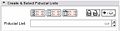 CreateSelectFiducialLists-3.4.jpg 375 × 98; 9 KB
 AddConfigureDeleteFiducials-3.4.jpg 364 × 59; 8 KB
 SlicerFiducialsAddNew-3.4.png 21 × 21; 271 bytes
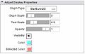 FiducialsAdjustDisplayProperties.jpg 361 × 231; 13 KB
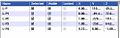 SlicerFiducialsListBox.jpg 493 × 155; 19 KB
 SlicerRealign-3.4.jpg 397 × 417; 31 KB
 EMSegment-Result.png 816 × 303; 213 KB
- Slicer3-4-v4cover.ppt ; 3.36 MB
 Slicer3.4.png 1,531 × 1,010; 862 KB
 Allapplication.png 1,920 × 1,150; 56 KB
 Allapplication scaled.png 1,736 × 1,040; 142 KB
 Allapplication scaled02.png 1,552 × 930; 123 KB
 Slicer3.4-2.png 1,972 × 1,375; 1.48 MB
 FidGUI3.4Draft1.png 576 × 708; 41 KB
- TrackerSimulator Darwin x86.tgz ; 49 KB
- CT-angio-04-2009.zip ; 111.64 MB
 Flowchart.JPG 800 × 600; 38 KB
- 58 IM-0009-0001.zip ; 3.7 MB
- 65 IM-0009-0001.zip ; 2.83 MB
- 57 IM-0009-0001.zip ; 2.93 MB
 FidGUIAnno.png 632 × 682; 71 KB
- IM-0004-0001.zip ; 3.65 MB
- 64 IM-0009-0001.zip ; 3.65 MB
 Z0.JPG 256 × 256; 10 KB
 1 Z0.JPG 256 × 256; 10 KB
 Z1.JPG 256 × 256; 14 KB
 MeanSliceZ.JPG 256 × 256; 10 KB
 64 58 Affine 150 Iterations.jpeg 1,918 × 1,200; 279 KB
 64 58 Affine 250 Iterations.jpeg 1,918 × 1,200; 275 KB
 64 58 Affine 350 Iterations.jpeg 1,918 × 1,200; 445 KB
 64 58 Affine 450 Iterations.jpeg 1,918 × 1,200; 461 KB
 64 58 Affine 500 Iterations.jpeg 1,918 × 1,200; 443 KB
- Slicer3minute.zip ; 15.93 MB
 MRI Bias Field Correction Before.png 664 × 655; 192 KB
 MRI Bias Field Correction After.png 664 × 656; 203 KB
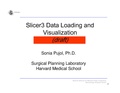 Slicer3Course DataLoadingAndVisualization SoniaPujol-DRAFT.pdf 1,650 × 1,275, 87 pages; 15.47 MB
 FourDAnalysisModuleScreenShot Prostate-3.5.png 800 × 499; 206 KB
 INRIAlogo.png 281 × 255; 23 KB
- Collidingfronts.swf ; 38.08 MB
- Tree.swf ; 32.53 MB
- Aneurysm.swf ; 40.66 MB
 VMTKLevelSetSegmentation Features.png 600 × 500; 162 KB
 VMTKVesselEnhancement gui.png 377 × 582; 17 KB
 VMTKVesselEnhancement process1.png 425 × 232; 42 KB
 VMTKVesselEnhancement process2.png 427 × 232; 49 KB
 VMTKVesselEnhancement io panel.png 645 × 187; 10 KB
 VMTKVesselEnhancement vesselenhancement panel.png 675 × 400; 19 KB
 ProgrammingIntoSlicer3 SoniaPujol-draft.pdf 1,650 × 1,275, 70 pages; 32.61 MB
 Slicer3.4-module-path-dialog.png 1,125 × 520; 40 KB
 VMTKEasyLevelSetGUI.png 381 × 884; 22 KB
 VMTKEasyLevelSetSegmentationAneurysm.png 552 × 445; 46 KB
- VMTKEasyLevelSetSegmentationAneurysm.swf ; 15.6 MB
 VMTKEasyLevelSetSegmentationBronchi.png 776 × 681; 423 KB
 VMTKEasyLevelSetSegmentationClassDiagram.png 961 × 888; 77 KB
 VMTKEasyLevelSetSegmentationCoronaries.png 511 × 528; 81 KB
 VMTKEasyLevelSetSegmentationEvolution.png 602 × 278; 23 KB
- VMTKEasyLevelSetSegmentationHeart.swf ; 14.77 MB
 VMTKEasyLevelSetSegmentationInitialization.png 597 × 109; 8 KB
 VMTKEasyLevelSetSegmentationInputOutput.png 660 × 307; 28 KB
- VMTKEasyLevelSetSegmentationLung.swf ; 20.51 MB
 VMTKEasyLevelSetSegmentationParameters.png 576 × 121; 13 KB
 VMTKEasyLevelSetSegmentationFM.png 1,156 × 621; 43 KB
 VMTKEasyLevelSetSegmentationGeodesic.png 1,135 × 660; 42 KB
 BCAnalysis Perfusion1.png 1,318 × 970; 296 KB
 Slicer3Course DataLoadingAndVisualization SoniaPujol.pdf 1,650 × 1,275, 111 pages; 26.29 MB
 Lupus3.png 164 × 202; 12 KB
 StochasticTractographyTutorial.png 340 × 236; 57 KB
 LupusTutorial.png 280 × 254; 33 KB
 800px-AdvancedNavigationTutorialSummary.png 800 × 500; 168 KB
 Slicer3-IGT-LEGOTutorial-biopsy.jpg 800 × 600; 62 KB
 AdvancedNavigationTutorialSummary.png 800 × 500; 168 KB
 Shot1.TIF 923 × 541; 122 KB
 Shot2.JPG 910 × 532; 219 KB
 Shot1.JPG 923 × 541; 79 KB
 Shot3.JPG 914 × 535; 79 KB
 Slicer VMTK student research project.pdf 1,275 × 1,650, 38 pages; 2.46 MB
 Multimuscle1.png 648 × 1,163; 753 KB
 Airways34.pdf 415 × 473; 48 KB
 Phillips-CJVR2009.pdf 1,218 × 1,631, 7 pages; 3.52 MB
 Walhovd-NeurobiolAging2009.pdf 1,239 × 1,654, 17 pages; 1.24 MB
 Fjell-NeuroImage2008.pdf 1,240 × 1,653, 15 pages; 4.16 MB
 PCN hayano.pdf 1,240 × 1,624, 11 pages; 295 KB
- Oct2009.zip ; 65.03 MB
 Cherbuin-PLoS-ONE2009.pdf 1,275 × 1,647, 10 pages; 672 KB
 MRMLPreset.png 1,200 × 794; 676 KB
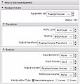
 QtSlicer-2009 10 02-QtTransformModule.png 1,160 × 916; 106 KB
 2009 10 02-QtTransformModule-v2.png 928 × 908; 64 KB
 2009 10 16-QCTKCollapsibleGroupBox.png 400 × 139; 3 KB
 2009 10 23-QCTKCollapsibleWidget.png 400 × 300; 7 KB
 2009 10 23-QCTKColorPickerButton.png 967 × 430; 34 KB
 2009 10 16-QCTKCoordinatesWidget.png 373 × 84; 5 KB
 2009 10 02-QCTKLinearTransformSlider.png 406 × 61; 3 KB
 2009 10 02-QMRMLNodeSelector.png 479 × 77; 4 KB
 2009 10 27-QCTKTitleComboBox.png 206 × 152; 5 KB
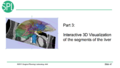
- 3DVisualization SoniaPujol RSNA2008.ppt ; 31.14 MB
- LiverSegmentation.zip ; 39.18 MB
 2009 11 10-qSlicerTransformsModuleUI.png 400 × 712; 32 KB
 2009 11 06-qCamerasModule.png 400 × 509; 8 KB
 Vmtkaftercenterlineonlyvoronoi.png 1,262 × 853; 83 KB
- Slicer3.4.1-ChangeTrackerTutorial.ppt ; 4.57 MB
 QCTKTreeComboBox.png 324 × 236; 10 KB
 Screenshot1.png 998 × 600; 1.71 MB
- Morphology.zip ; 33.6 MB
 BrainH1.png 808 × 827; 538 KB
 Bumps conv 1.png 804 × 831; 180 KB
 Curvature bumps1 good.png 722 × 634; 348 KB
 Sulci3 conv 2.PNG 158 × 168; 36 KB
 Sulci3 init 3.png 632 × 672; 193 KB
 Sulci6 done2.png 161 × 188; 32 KB
 3DVisualization SoniaPujol RSNA2009 11-30-2009.pdf 1,500 × 1,125, 125 pages; 22.22 MB
- LiverData.zip ; 502 bytes
- LiverData.tar.gz ; 33.05 MB
 Vmtkcenterlinesgui.png 396 × 562; 18 KB
- Slicer3 Tutorial ManualRegistration.ppt ; 3.27 MB
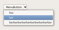 QCTKMenuButton.png 291 × 149; 4 KB
- Demo1.zip ; 119.34 MB
 Eclipse-build-options.png 999 × 558; 93 KB
 QCTKDoubleSlider.png 259 × 47; 932 bytes
 QCTKSliderSpinBoxWidget.png 244 × 38; 2 KB
 QCTKFittedTextBrowser.png 663 × 398; 49 KB
 QCTKFittedTextBrowser2.png 363 × 632; 43 KB
- Stochastic June09 2.ppt ; 9.33 MB
 QCTKRangeSlider.png 228 × 39; 977 bytes
 Registration Demons icon.png 200 × 200; 21 KB
 CtkDICOMModel.png 963 × 734; 88 KB
 QCTKRangeWidget.png 310 × 46; 2 KB
 Logo mimx.gif 107 × 81; 1 KB
 Slicer3-Brainlab-Connect.png 1,496 × 949; 50 KB
 VTK-CPU.png 547 × 490; 202 KB
 VTK-GPU.png 547 × 487; 236 KB
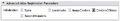 ARCTIC AdvancedAtlasRegParam.png 431 × 75; 2 KB
 ARCTIC AdvancedCortThickParam.png 432 × 76; 1 KB
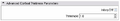
 ARCTIC AdvancedSkullStrippingParam.png 433 × 53; 1 KB
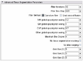 ARCTIC AdvancedTissueSegParam.png 431 × 320; 5 KB
 CartilegeMIdGEMRIC15T.png 52 × 21; 6 KB
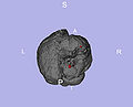 LesionInBrain.jpg 864 × 699; 109 KB
 RSS MultiObjSeg1.png 852 × 602; 240 KB
 VtkGPURayCasting-AutoClipping.png 1,280 × 754; 274 KB
 VtkGPURayCasting-AutoClipping-CPU.png 1,280 × 754; 301 KB
 VtkGPURayCasting-DualView.png 1,204 × 972; 277 KB
 QCTKCheckableHeaderView.png 240 × 138; 6 KB
 ResultSubtractImage.png 320 × 147; 28 KB
 VR-GUI.png 458 × 698; 14 KB
 CtkTransferFunctionWidget.png 651 × 162; 8 KB
- SampleData Grayscale.nrrd ; 19.88 MB
 CtkDirectoryButton.png 291 × 55; 4 KB
 DicomToNrrd-IOPanels-3-6.png 395 × 195; 8 KB
 Slicer3.6MultiplyImage-GUI.jpg 399 × 334; 22 KB
 MITLogo.png 62 × 36; 184 bytes
 NAMICLogo.png 33 × 40; 2 KB
 SPLLogo.png 100 × 100; 10 KB
 Training-2010-05-02.png 1,269 × 848; 310 KB
 Slicer3 brainlabmodule load may3.png 1,024 × 768; 151 KB
 Slicer3 brainlabmodule connect may3.png 1,024 × 768; 153 KB
 Slicer3 brainlabmodule navigate may3.png 1,024 × 768; 152 KB
- DTI-Brain.nrrd ; 7.39 MB
- CT-chest.nrrd ; 40.23 MB
- MR-head.nrrd ; 6.3 MB
 Mask.png 843 × 753; 216 KB
- RegLib C01 1.nrrd ; 4.83 MB
- RegLib C01 2.nrrd ; 5.85 MB
- CTA-cardio.nrrd ; 61.08 MB
 Slicer36-LayoutMenu.png 606 × 374; 26 KB
 BuildInstructions-Slicer-UseQt.png 857 × 640; 47 KB
 BuildInstructions-Slicer-CTK DIR.png 408 × 234; 8 KB
 BuildInstructions-CTK-build-VTK DIR.png 408 × 234; 8 KB
 BuildInstruction-CTK-build.png 857 × 640; 43 KB
 Slicer3-6icons.png 647 × 344; 447 KB
 BRAINSFitUI.png 373 × 714; 24 KB
 New hellopython image 3.6tutorial.png 628 × 335; 95 KB
 HelloPython.png 692 × 297; 113 KB
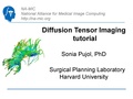
 DiffusionMRITutorial Slicer3.6 SPujol.pdf 1,500 × 1,125, 66 pages; 5.18 MB
 Slicer3.5GyriContourSegmentation.png 384 × 729; 19 KB
 Slicer3-BRAINSMushExtractionUtilityGUI.png 384 × 729; 19 KB
 LungVolumeRenderingUnfiltered.png 1,213 × 912; 1.74 MB
 LungVolumeRenderingFiltered.png 1,206 × 957; 1.54 MB
 Bennett-PLoSPathogens2010.pdf 1,275 × 1,647, 10 pages; 1.69 MB
 Rigid reg T1-Scan.gif 196 × 215; 37 KB
 Rigid reg T1-Scan b0 overlay.gif 196 × 215; 27 KB
 DiffusionMRITutorial Slicer3.6 SoniaPujol.pdf 1,500 × 1,125, 66 pages; 5.19 MB
 Slicer3.6MinuteTutorial SoniaPujol.pdf 1,500 × 1,125, 32 pages; 2.28 MB
 IAFEMesh-TutorialContestSummer2010.pdf 1,500 × 1,125, 47 pages; 1.71 MB
 Fiducials TutorialContestSummer2010.pdf 1,500 × 1,125, 98 pages; 2.4 MB
 Longitudinal Lesion Comparison TutorialContest 2010.pdf 1,500 × 1,125, 32 pages; 662 KB
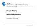
 Slicer3.6 Tutorial ManualRegistration.pdf 1,500 × 1,125, 14 pages; 1.11 MB
 3DDataLoadingAndVisualization Slicer3.6 SoniaPujol.pdf 1,500 × 1,125, 111 pages; 8.2 MB
 ProstateNav TutorialContestSummer2010.pdf 1,500 × 1,125, 29 pages; 2.54 MB
 LabelFusion Tutorial.pdf 1,650 × 1,275, 34 pages; 1.23 MB
- Labelmap Seeding Model.vtk ; 8.22 MB
- Labelmap Seeding Model Double.vtk ; 8.1 MB
 InteractiveEditorTutorial SoniaPujol.pdf 1,500 × 1,125, 61 pages; 6.35 MB
 QMRMLSliceViewWidget.png 348 × 428; 39 KB
 Logo-LMI.png 100 × 119; 18 KB
 SlicerLoadScene.png 107 × 111; 3 KB
 DiffusionMRITutorial Slicer3.6.1 SoniaPujol Aug2010.pdf 1,500 × 1,125, 68 pages; 7.99 MB
 ManualRegistration 3.6 noGIF.pdf 1,654 × 1,239, 14 pages; 1.08 MB
- ManualRegistration Slicer3.6.ppt ; 1.58 MB
- ProgrammingIntoSlicer3.6 SoniaPujol.ppt ; 5.01 MB
 Jolley-HR2008.pdf 1,275 × 1,650, 19 pages; 1.24 MB
 Lindig-HistochemCellBiol2009.pdf 1,275 × 1,650, 21 pages; 2.01 MB
 Lindig-HistochemCellBiol2009-fig7.png 1,200 × 496; 232 KB
 ProgrammingIntoSlicer3.6 Python SoniaPujol.pdf 1,500 × 1,125, 52 pages; 3.17 MB
 QSlicerScalarVolumeDisplayWidget.png 433 × 495; 33 KB
 QMRMLColorTableNodeComboBox.png 205 × 685; 16 KB
 Liao-IEEE-TBME2010-fig1.png 2,000 × 1,067; 1.35 MB
 Liao-IEEE-TBME2010.pdf 1,237 × 1,650, 12 pages; 1.36 MB
- SlicerNeurosurgeryTutorial-3.6.ppt ; 12.54 MB
- SlicerNeurosurgeryTutorial-3.6.1.ppt ; 10.41 MB
- Slicer-tutorial-neurosurgery.zip ; 78.28 MB
 Slicer3-6Announcement-v1.png 216 × 144; 51 KB
 InteractiveEditorTutorial Slicer3.6 SoniaPujol.pdf 1,500 × 1,166, 61 pages; 10.69 MB
 PythonTutorial.PNG 1,047 × 740; 237 KB
 RSS TutorialContestSummer2010.pdf 1,500 × 1,125, 29 pages; 1.35 MB
 Stochastic Tractography TutorialContestSummer2010.pdf 1,500 × 1,125, 40 pages; 957 KB
- CASE42.nrrd ; 5.05 MB
- Avf voismall.nrrd ; 144 KB
 EMSegmenterTutorialAdvancedMode-2010-Nov.pdf 1,654 × 1,239, 59 pages; 7.97 MB
 EMSegmenterTutorialSimpleMode-2010-Nov.pdf 1,654 × 1,239, 16 pages; 1.71 MB
 QMRMLColorPickerWidget.png 300 × 280; 10 KB
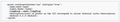
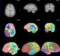
 MRI-Human-Brain-Parcellation T1 3x360x360.png 1,080 × 360; 135 KB
 MRI-Human-Brain-Parcellation 3x360x360.png 1,080 × 360; 25 KB
 MRI-Human-Brain-Parcellation.png 1,260 × 420; 241 KB
 DiffusionMRITutorial Slicer3.6.1 SoniaPujol Dec2010.pdf 1,500 × 1,125, 71 pages; 8.19 MB
- Slicer3Minute SoniaPujol 3.6.1.ppt ; 3.05 MB
- Slicer3.6-ChangeTrackerTutorial.ppt ; 5.41 MB
 Slicer3 DataLoadingAndVisualization SoniaPujol3.6.pdf 1,500 × 1,125, 107 pages; 8.12 MB
 SEM snippet.png 600 × 109; 8 KB
 EM-Segmenter.png 1,542 × 1,451; 489 KB
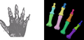 CTHandBone BeforeAfter.png 692 × 332; 154 KB
 Ac gui.png 404 × 760; 32 KB
 Afrer reg.png 510 × 510; 87 KB
 Midas-slicer-screenshot-upload3.jpg 1,185 × 753; 468 KB
 Star-forming.png 579 × 580; 348 KB
 ACcombined.png 1,840 × 577; 40 KB
 EMSegmentRev PETCT A.png 437 × 522; 87 KB
 EMSegmentRev PETCT B.png 433 × 522; 115 KB
 EMSegmentRev PETCT C.png 434 × 521; 79 KB
 EMS Hemisphere labelmap50.png 1,292 × 463; 450 KB
- Error creating thumbnail: File with dimensions greater than 12.5 MPSlicer rdep squeeze.png 12,532 × 2,685; 6.19 MB
 Slicer4HelpExample.png 420 × 383; 13 KB
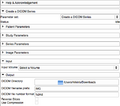
 Slicer4AcknowledgementExample.png 420 × 377; 45 KB
 SliceViewers-Hi3.png 674 × 355; 45 KB
 Slicer-Conventional-2011-7-32-mac.png 1,115 × 466; 38 KB
 Slicer36 compendium.png 836 × 751; 263 KB
 EndoscopyGUI.png 441 × 445; 23 KB
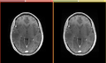 GradientAnisotropicDiffusion-Before-After-2011-08-11.png 932 × 560; 225 KB
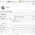
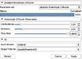 GradientAnisotropicDiffusion-Panel-2011-08-11.png 439 × 317; 33 KB
 ImageReadDicomWrite-2011-08-26.png 516 × 455; 44 KB
 3D Slicer 4.0.gamma-2011-10-20 136.png 1,484 × 1,031; 640 KB
 Selection 140.png 997 × 503; 77 KB
 Slicer4-SceneViewCreateEdit.jpeg 469 × 447; 29 KB
 NCIGT logo.gif 128 × 128; 4 KB
 UIowa-logo.jpg 192 × 174; 34 KB
 ComputeSUVBodyWeightUI.png 576 × 385; 39 KB
 Slicer4minute-tutorial SoniaPujol.pdf 1,500 × 1,125, 25 pages; 8.79 MB
 MainGUI-2011-11-24.png 1,741 × 1,337; 132 KB
 SlicerApplication-AddVolume.png 607 × 449; 26 KB
 SlicerApplication-FileAddVolume.png 220 × 286; 14 KB
 SlicerApplication-AddVolume-SlicerWelcome.png 268 × 156; 7 KB
 SlicerApplication-AddVolume-SingleFile.png 607 × 449; 31 KB
 Slicer4-Mask-GUI.jpeg 585 × 807; 62 KB
 Slicer4Announcement-HiRes crop.png 525 × 312; 213 KB
 Slicer4RSNA.png 1,792 × 1,008; 613 KB
 SlicerApplication-SaveData-SelectSceneAndData.png 32 × 32; 2 KB
 SlicerApplication-SaveData-SelectModifiedData.png 32 × 32; 1 KB
 Berman-JNeurosurg2004.pdf 1,218 × 1,631, 7 pages; 255 KB
 Lewis-CerebCortex2009.pdf 1,237 × 1,631, 9 pages; 651 KB
 Majewicz-IEEEIntConfRobotAutom2010.pdf 1,275 × 1,650, 20 pages; 735 KB
 Caskey-journal.pone.0027372-fig3.png 1,174 × 439; 321 KB
 SlicerApplication-AddData-SlicerWelcome.png 490 × 292; 42 KB
 LoadSaveToolbar-2012-02-22 at 4.28.51 PM.png 124 × 40; 7 KB
 Slicer-r19441-CLIExtensionTemplate-screenshot.png 679 × 589; 49 KB
 Slicer-r19441-LoadableExtensionTemplate-screenshot.png 610 × 430; 47 KB
 Slicer-r19441-ScriptedLoadableExtensionTemplate-screenshot.png 620 × 571; 72 KB
 Slicer-r19441-SuperBuildLoadableExtensionTemplate-screenshot.png 629 × 513; 51 KB
 Slicer-r19441-CLIExtensionTemplate-screenshot-2.png 801 × 561; 80 KB
 WebinarSlicer40.png 640 × 400; 136 KB
 TransformsModuleTransformedNodes.png 437 × 530; 15 KB
 Plastimatch.png 335 × 335; 7 KB
 UIowaDome-Small.gif 21 × 32; 433 bytes
 ScreenShot GoogleHangout.PNG 518 × 478; 21 KB
 QtTestingRecorderMenu.jpg 454 × 154; 26 KB
- 3DSlcierQtTesting-0001.mpeg ; 4.35 MB
 PkModelingUI.png 447 × 726; 33 KB
 QtTestintMacroFileDialog.jpg 536 × 325; 58 KB
 PkModelingUI061212.png 447 × 617; 28 KB
 SlicerDeveloper-ContributionWorkflow.png 1,771 × 755; 133 KB
 Play.png 150 × 150; 21 KB
 Wand-icon-extension.png 439 × 212; 21 KB
 SlicerRtExtensionLogo.png 128 × 128; 24 KB
 3DSlicerQtTesting.gif 400 × 240; 16.09 MB
- TestData mrb.zip ; 46.19 MB
- TestData MRB format.zip ; 57.79 MB
 PerkNavLogo.png 128 × 128; 25 KB
 PerkNavScreenshot.png 1,565 × 1,050; 251 KB
- Dti-cc.zip ; 42.31 MB
 IGyne 1.0 needle visualization.png 2,560 × 1,440; 1.78 MB
 IGyne 1.0-registration.png 2,560 × 1,440; 1.24 MB
 Igyne 1.0-Tumor and Needle Visualization.png 2,560 × 1,440; 1.39 MB
 Igyne Icon.png 128 × 128; 20 KB
- Igyne Tutorial.ppt ; 6.5 MB
- IGyne1.0 Tutorial.ppt ; 6.5 MB
 SOV Icon 128.png 128 × 128; 30 KB
 Spatialobjects icon.png 128 × 128; 7 KB
 Spatial objects screenshot.png 800 × 600; 245 KB
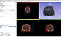 LongitudinalPETCT Screenshot.jpg 1,920 × 1,175; 233 KB
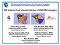
 3DVisualizationDICOM RadiologyApplications SoniaPujol KittShaffer RSNA2012.pdf 1,500 × 843, 107 pages; 49.98 MB


